

TracIcon
Retrospective motion correction for 3D brain MRI. Use your same protocol - with no change to the acquired data. Motion happens, TracIcon corrects.
Pending regulatory clearances. The product is pending CE certification in Europe. The product is not cleared or approved by the FDA and is not available for sale in the United States.
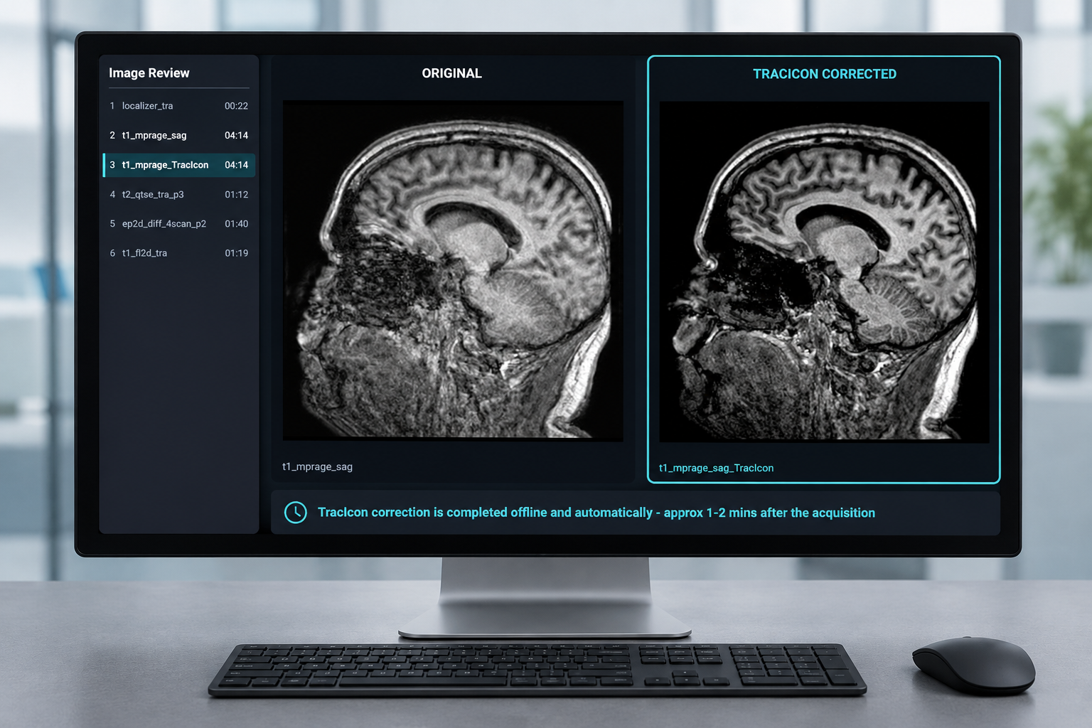
TracPoint-informed retrospective motion correction for brain MRI.
Motion in a 3D dataset forces a difficult choice: repeat the sequence, accept a degraded image, switch to a less suitable or faster low resolution sequence, or sedate the patient.
TracIcon can enable you to recover motion degraded 3D imaging without always needing to rescan. Correction is performed automatically for compatible datasets using optical tracking data from TracPoint, with head position changes measured 30 times per second.
How it works
TracIcon makes motion in 3D dataset a correctable event, not an unrecoverable failure. Run the sequence with TracIcon active and receive a motion-corrected series automatically. The corrected images can be presented side-by-side with the original for review.

- 1
Calibrate
A short 15-second calibration scan aligns the spatial coordinate system of the MRI scanner to the coordinate system of TracPoint.
- 2
Track
TracPoint records precise 6-DoF head motion throughout the scan.
- 3
Synchronize
Motion data is time-matched to the moment each part of the image was acquired.
- 4
Correct
TracIcon corrects the motion-affected data after acquisition. Correction is performed in under a minute.
- 5
Review
A corrected series returns to the console alongside the original, ready to read.
Why it's different
Works with your sequence
Compatible sequences run exactly as designed. A short calibration aligns TracPoint to the MR scanner imaging coordinates.
Precise optical input
Driven by the precise optical tracking measured 30 times per second. No AI, no machine learning. Never estimated from the image itself.
Automatic correction
Runs on the TracSuite workstation and in the background, with minimal operator input. Pre-requisite is TracSuite and TracPoint.
Multi-vendor
Currently 3D-GRE and 3D-TSE sequences from Siemens and Philips MR scanners are supported.
Before and after
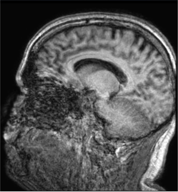
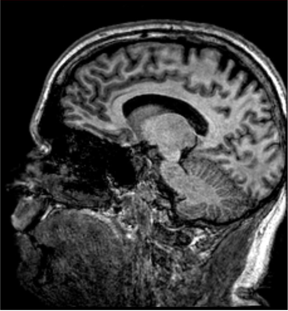
Volunteer imaging acquired on Philips Achieva 3T. Courtesy of Glostrup Hospital, Rigshospitalet, Copenhagen.
WHO IT'S FOR
Completes the chain

HARDWARE
Monitor live.
The markerless optical hardware that records the motion data TracIcon uses for correction.

Operating Software
Monitor and Inform.
The operating software for TracPoint, showing motion at the console and keeping the motion record. Pre-requisite for TracIcon.